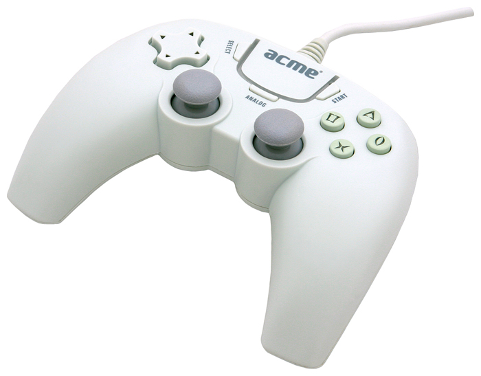
Hansmann Joystick Driver

Apr 15, 2013 - Download Joystick Driver for Java for free. A Java interface to the joystick or any other input device with 2-6 degrees of freedom (Works on.
Chemical kinetics and reaction mechanisms espenson pdf the best free software f. A medical imaging and therapy device is provided that may include any of a number of features. One feature of the device is that it can image a target tissue volume and apply ultrasound energy to the target tissue volume.
In some embodiments, the medical imaging and therapy device is configured controllably apply ultrasound energy into the prostate by maintaining a cavitational bubble cloud generated by an ultrasound therapy system within an image of the prostate generated by an imaging system. The medical imaging and therapy device can be used in therapeutic applications such as Histotripsy, Lithotripsy, and HIFU, for example. Methods associated with use of the medical imaging and therapy device are also covered. CROSS REFERENCE TO RELATED APPLICATIONS This application claims the benefit under 35 U.S.C.
Provisional Patent Application No. 61/237,017, filed Aug. 26, 2009, titled “MICROMANIPULATOR CONTROL ARM FOR THERAPEUTIC AND IMAGING ULTRASOUND TRANSDUCERS”. This application is herein incorporated by reference in its entirety. INCORPORATION BY REFERENCE All publications, including patents and patent applications, mentioned in this specification are herein incorporated by reference in their entirety to the same extent as if each individual publication was specifically and individually indicated to be incorporated by reference. FIELD OF THE INVENTION The present invention generally relates to imaging and treating tissue with ultrasound devices. More specifically, the present invention relates to imaging and ablating tissue with Histotripsy devices.

BACKGROUND OF THE INVENTION Histotripsy and Lithotripsy are non-invasive tissue ablation modalities that focus pulsed ultrasound from outside the body to a target tissue inside the body. Histotripsy mechanically damages tissue through cavitation of micro bubbles which homogenizes cellular tissues into an a-cellular liquid that can be expelled or absorbed by the body, and Lithotripsy is typically used to fragment urinary stones with acoustic shockwaves. Read my lips the balm. Histotripsy is the mechanical disruption via acoustic cavitation of a target tissue volume or tissue embedded inclusion as part of a surgical or other therapeutic procedure. Histotripsy works best when a whole set of acoustic and transducer scan parameters controlling the spatial extent of periodic cavitation events are within a rather narrow range. Small changes in any of the parameters can result in discontinuation of the ongoing process. Histotripsy requires high peak intensity acoustic pulses which in turn require large surface area focused transducers. These transducers are often very similar to the transducers used for Lithotripsy and often operate in the same frequency range.
The primary difference is in how the devices are driven electrically. Histotripsy pulses consist of a (usually) small number of cycles of a sinusoidal driving voltage whereas Lithotripsy is (most usually) driven by a single high voltage pulse with the transducer responding at its natural frequencies. Even though the Lithotripsy pulse is only one cycle, its negative pressure phase length is equal to or greater than the entire length of the Histotripsy pulse, lasting tens of microseconds. This negative pressure phase allows generation and continual growth of the bubbles, resulting in bubbles of sizes up to 1 mm. The Lithotripsy pulses use the mechanical stress produced by a shockwave and these 1 mm bubbles to cause tissue damage or fractionate stones. In comparison, each negative and positive cycle of a Histotripsy pulse grows and collapses the bubbles, and the next cycle repeats the same process. The maximal sizes of bubbles reach approximately tens to hundreds of microns.